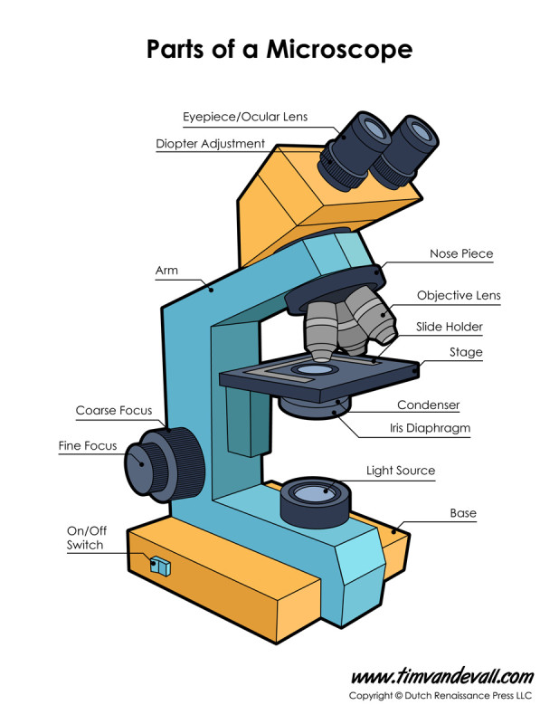
Have you ever wondered how scientists are able to peer into the intricate world of cells, bacteria, and other microscopic wonders? The answer lies in a crucial component found within microscopes, the diaphragm. This seemingly simple device plays a vital role in sharpening the images we see, allowing us to explore the unseen realms that shape our world.

Image: byjus.com
The diaphragm, a circular aperture with adjustable diameter, lies at the heart of how light is controlled in a microscope. But beyond its simple appearance, the diaphragm unveils a complex interplay of light and optics, enabling users to fine-tune the illumination and contrast of their microscopic observations. In this article, we delve into the fascinating world of the diaphragm, exploring its structure, function, and how it dramatically impacts the clarity and detail revealed by microscopes.
Understanding the Basics of Microscope Illumination
To understand the role of the diaphragm, it’s essential to grasp the fundamental principles of light in microscopy. Microscopes work by illuminating the object being examined with light, which then passes through lenses to magnify the image. The path and intensity of this light are crucial in producing a clear and detailed view.
There are two primary types of illumination used in microscopy:
- Transmitted Illumination: The light source is positioned beneath the specimen, so the light travels through the object before reaching the objective lens.
- Reflected Illumination: The light source is positioned above the specimen, and the light is reflected off the surface of the object before reaching the objective lens.
The Diaphragm’s Role in Light Control
The diaphragm acts like a gatekeeper, controlling the amount of light that reaches the objective lens. It’s located within the condenser, a component positioned below the stage of the microscope, which focuses the light onto the specimen.
Adjusting Light Intensity
By opening and closing the diaphragm, you can increase or decrease the light intensity. Opening the diaphragm allows more light to pass through, while closing it restricts the light flow. This control over light intensity is critical for achieving optimal illumination of the specimen, depending on its transparency and the desired level of detail.

Image: priaxon.com
Controlling Contrast and Depth of Field
Apart from light intensity, the diaphragm also influences the contrast and depth of field in the image.
Here’s a breakdown:
- Contrast: The diaphragm controls the amount of stray light, which can create unwanted glare and blur the image. By closing the diaphragm, you reduce stray light, enhancing the contrast of the image and making details more visible.
- Depth of Field: The diaphragm impacts the depth of field, the area of the specimen that appears in sharp focus. With a closed diaphragm, depth of field is increased, meaning a larger portion of the specimen remains in focus. Conversely, opening the diaphragm decreases depth of field, focusing on a thinner layer of the specimen. This ability to control depth of field is particularly valuable in observing thick specimens or those with intricate structures.
Types of Diaphragms
There are several types of diaphragms employed in microscopes, each offering different levels of control and application:
1. Iris Diaphragm
The most common type of diaphragm, the iris diaphragm, resembles the iris of the human eye. It consists of a set of overlapping metal blades that can be opened or closed using a lever. This design provides a precise and continuous adjustment of the light aperture.
2. Disc Diaphragm
Disc diaphragms, simpler in design, use a series of punched metal discs with different aperture sizes. This type offers less precise adjustment compared to the iris diaphragm.
3. Aperture Diaphragm
The aperture diaphragm is located in the condenser, directly beneath the stage. It controls the size of the light cone that illuminates the specimen. Its primary function is to optimize the illumination quality for different magnification levels.
4. Field Diaphragm
Unlike the aperture diaphragm, the field diaphragm is located below the light source. It controls the overall diameter of the light beam that reaches the condenser. By adjusting the field diaphragm, you can limit the area of the specimen illuminated, preventing stray light and improving image clarity.
Diaphragm Adjustment for Optimal Imaging
Mastering the art of diaphragm adjustment is crucial for achieving optimal image quality in microscopes. It’s a process of balancing light intensity, contrast, and depth of field to reveal the most intricate details of your specimen.
Here’s a general guide to diaphragm adjustment:
- Start with a fully opened diaphragm: This allows maximum light to pass through, providing a bright and clear view.
- Observe the image: Carefully assess the specimen’s details and illumination.
- Adjust the diaphragm: Gently close the diaphragm to:
- Reduce glare: If the image is too bright or exhibits excessive glare, closing the diaphragm will improve contrast.
- Improve the resolution: Closing the diaphragm can enhance the sharpness and clarity of the image.
- Focus on specific areas: Minimizing the depth of field by closing the diaphragm can help you focus on specific details within a complex specimen.
- Fine-tune for optimal results: Continue adjusting the diaphragm until you achieve the desired level of light intensity, contrast, and depth of field.
Beyond Microscopy: Applications of Diaphragms
While diaphragms are essential for microscope function, they see use in many other fields as well:
Here are some examples:
- Photography: Diaphragms are integral to cameras, controlling the amount of light that reaches the camera sensor and influencing the depth of field in images.
- Telescopes: Telescopes use diaphragms to regulate light flow and enhance image quality, especially when observing faint celestial objects.
- Other scientific instruments: Diaphragms are often used in laser systems, spectrometers, and other instruments to modulate and control the intensity of light beams.
Diaphragm Microscope Function
The Future of Diaphragms in Microscopy
Research and innovation continue to shape the future of microscopy and the role of diaphragms within it. Recent advancements in light sheet microscopy, for instance, use ultra-thin sheets of light to illuminate and image specimens with remarkable speed and detail. This innovative technique is revolutionizing 3D imaging in biological research and relies on precise diaphragm control for optimal results.
Furthermore, the integration of computer-controlled diaphragms and automated imaging systems is creating new possibilities for microscopists. This automation allows for greater precision, reproducibility, and efficiency in imaging complex specimens, opening doors to new discoveries and insights in various scientific fields.
The diaphragm, often unassuming yet crucial, will continue to play a critical role in enhancing our understanding of the microscopic world. Its ability to shape the flow of light and influence image quality makes it an indispensable tool for scientists, researchers, and enthusiasts alike. As our quest to unravel the secrets of tiny worlds continues, the diaphragm will remain a vital component, guiding us towards clearer and more detailed views of the universe around us.


